
Mr Josh Abram
Trainee Research Technician
QMUL ARC, Queen Mary University of London
Optimising a Human-Specific Lymph Node Model to Investigate EMT-Driven Metastasis in Oral Squamous Cell Carcinoma
Dr Adrian Biddle’s team uses advanced cell culture methods, organ-on-chip technology, and artificial intelligence (AI) to study oral cancer metastasis. With support from an ARC grant, Dr Biddle has appointed Josh Abram, a university graduate, as a research technician trainee to continue important work optimising a peptide hydrogel model of the lymph node.
The Need for Human-Specific Models to Study Oral Cancer Metastasis
Oral Squamous Cell Carcinoma (OSCC) is the most common mouth cancer. In around 50% of patients, the disease spreads (metastasises) to nearby lymph nodes – a development that is strongly linked to poorer clinical outcomes and reduced survival rate. Historically, studying this metastasis has relied heavily on the use of in vivo mouse studies, however, the translational applicability to patients is low as crucial aspects of the tumour microenvironment and immune component are lost in these models. To address this problem, Dr Biddle’s group are developing a more human-relevant peptide hydrogel model to study oral cancer metastasis.
What are Peptide Hydrogels and Why are they Exciting?
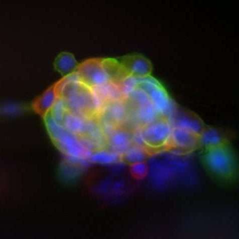
Peptide hydrogels are formed of short peptide repeats of the FEFEFKFK peptide that self-assemble to form stable nanofiber networks. These gels are optically clear and degradable, which allows fluorescent imaging of OSCC cells within the 3D matrix and retrieval of cells for characterisation via flow cytometry. Furthermore, the addition of different extracellular matrix (ECM) fibres to the gels enables the accurate mimicking of the types of 3D tissue interactions OSCC undergoes during lymph node metastasis.
Optimising a Peptide Hydrogel Model of the Lymph Node
Josh’s research continues from the work of final-year PhD student Momita Bhuiyan, who built and optimised a peptide hydrogel model of the lymph node. The model is a 3D biomimetic model, in which OSCC cells can be inserted and assessed for mechanisms of invasion. In this model foetal bovine serum (FBS) has been replaced with human platelet lysate (HPL), providing equal nutrient-dense supplementation, but specific for human cell culture. All proteins added to cell culture, or to the gels directly, are human-derived, ensuring the model is as relevant to patients as possible.
The Problems with FBS
FBS is a growth supplement produced by bovine foetuses as a byproduct of the meat industry. While it has long been used to support growth of cells in vitro, there are significant scientific and ethical drawbacks in its use. These include:
- The presence of non-human molecules can alter human cell behaviour, thereby reducing translational relevance to human disease research
- Large batch to batch variability in its composition, hence reproducibility of experiments is impaired
- Contamination with bacteria and viruses
Studying Epithelial-to-Mesenchymal Transition
Given that OSCC is an epithelial cancer, the area of interest in terms of metastatic invasion is the plastic and reversible process of Epithelial-to-Mesenchymal transition (EMT). The loss of epithelial apical-basal cell polarity and cell-cell adhesions is essential for cells to adopt mesenchymal phenotypes, where migratory capacity is drastically increased. This allows for subpopulations of OSCCs to leave the primary tumour and metastasise to distant tissues. The successful seeding of these OSCC subpopulations in the lymph node is not fully understood. Is it performed by EMT cells, or by more plastic cell populations? By imaging OSCCs during invasion within their peptide hydrogel model, and analysing cell population dynamics with distinct ECM components, Josh aims to better understand the exact conditions driving successful seeding of OSCC within the lymph node tissue.
Long Term Aims
The long-term aim of this project is to improve the model to fully mimic the lymph nodes subcapsular sinus, the physical barrier between the lymph circulation and the lymph organ. The first step will be to produce immortalised human primary lymphatic endothelial cells (LECs) via retroviral transduction and utilise the cells to form monolayers on top of the peptide gel. This will mimic the LEC monolayer of the subcapsular sinus and will allow further elucidation as to how communication between OSCCs and floor LECs influence OSCC metastatic seeding.
Quotes
Mr Josh Abram
Trainee Research Technician
QMUL ARC, Queen Mary University of London
“Creating models built entirely from human cells and human-derived materials can give us information that is more accurate and directly applicable to patient care.”

Dr Ross Dobie
Centre for Human Specific Research
“This project represents exactly the kind of forward-thinking, human-focused research we are proud to support – advancing the development of innovative human-specific technologies to better understand and ultimately improve outcomes for patients with oral cancer.”