

The Centre for Human Specific Research is working with the Spatial Biology team at Concept Life Sciences to validate two antibody mimetics to replace animal-derived antibodies in automated immunohistochemistry staining. In this project update (1 of 2), we share the outcomes from the first two phases of the Transition Consultancy which cover our initial consultation and recommendations.
Consultation
At Concept Life Sciences, the Spatial Biology team employs a range of techniques, including immunohistochemistry (IHC), to understand how drugs interact with and influence complex tissue environments. A critical component of this work is the use of antibodies, and the team utilises a validated catalogue that includes monoclonal, polyclonal, and recombinant options. For this Transition Consultancy project, we focused on replacing both recombinant antibodies and non-recombinant antibodies that require the use of animals or animal-derived products (ADPs) in their production.
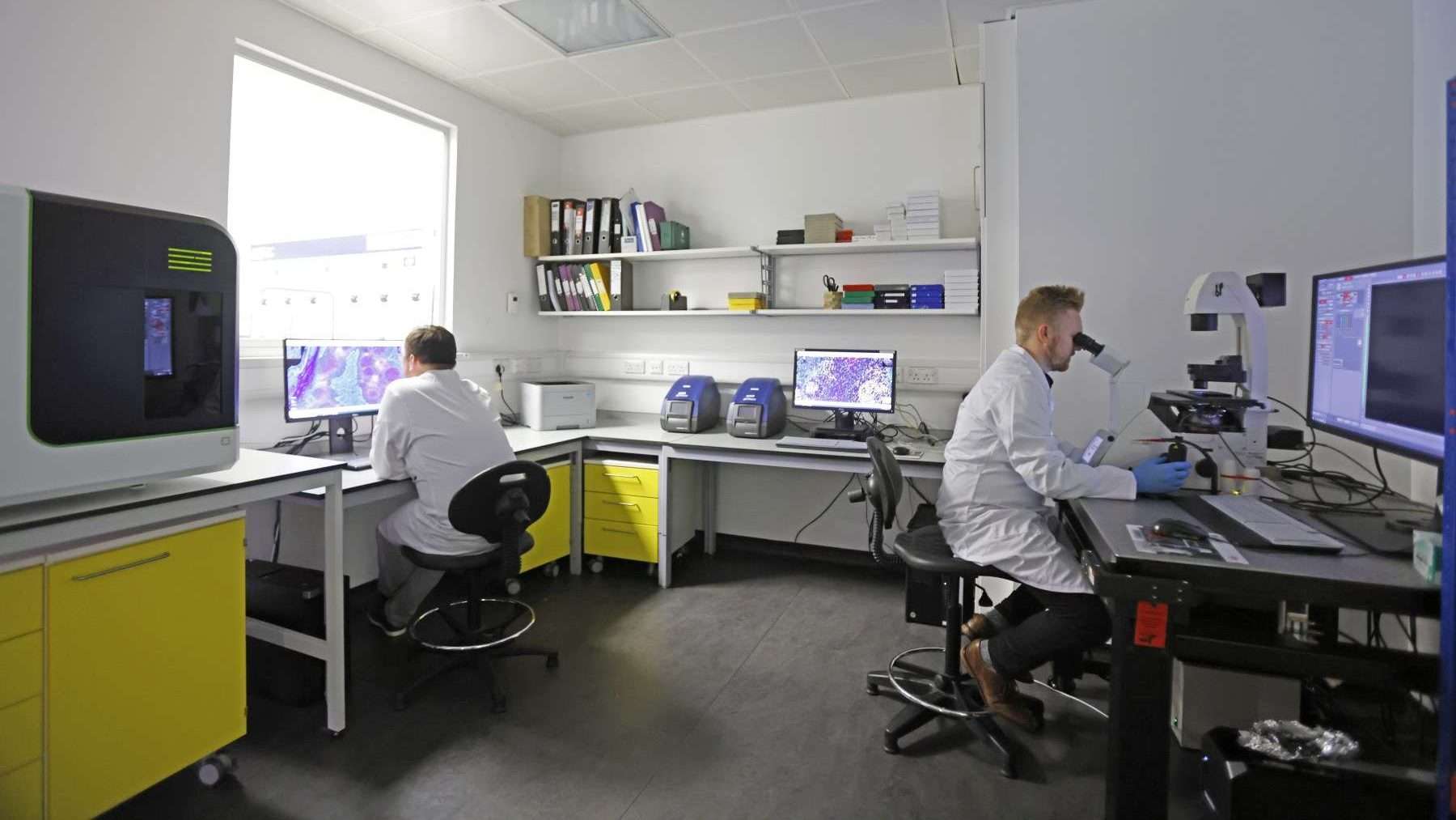
During our initial discussions, the team shared the challenges they have faced when using certain antibodies to stain diseased tissue samples. The most frequent issues encountered included poor staining quality and high background signal, both of which can hinder data interpretation. These recurring difficulties have prompted a growing interest in the team to investigate alternative staining strategies. Two particularly challenging targets were highlighted: CD4, a marker of T helper cells, and PD-L1, a key immune checkpoint protein. Achieving consistent and specific staining for these targets in diseased tissues has proven difficult with existing antibodies.
To address these issues, we explored the potential of antibody mimetics for IHC applications. Mimetics offer several advantages over antibodies, including the ability to circumvent common antibody-related limitations, such as high background. They can also be developed without relying on animals or ADPs, removing ethical concerns and reducing manufacturing issues such as batch-to-batch variation. Although the team have not previously worked with mimetics, they expressed strong interest in evaluating a new affinity reagent which could be easily integrated into their existing workflow and provide a novel solution for the staining of challenging tissues.
What are Antibody Mimetics?
An antibody mimetic is a type of molecule that mimics the ability of an antibody to bind specifically to a target (such as a protein, peptide, or small molecule), but it is not derived from immunoglobulin genes. In other words, it functions like an antibody but has a completely different structure.
By investigating antibody mimetics as viable alternatives, this project aims to address both the technical challenges of IHC in complex tissues and the ethical considerations surrounding the use of animal-based reagents. In addition, validating alternative staining approaches could provide significant value to the wider research community, extending benefits well beyond the immediate work at Concept Life Sciences.
Advise
Using the Recombinant Antibodies and Mimetics Database we were able to identify suitable CD4 and PD-L1 mimetics developed by ProImmune, known as Ankyrons. These small, single-domain binding reagents are a subtype of mimetic, based on an ankyrin-repeat scaffold. The Ankyrons listed in the database fall under Category A – which indicates they are completely animal-free. To learn more about mimetics, such as Ankyrons, visit the Production Methods section of our database.
An additional advantage of these affinity reagents lies in their simplified protocol. Because of their molecular design, no blocking step is required before application – removing the need for traditional blocking solutions, which typically contain animal-derived components. This not only removes another ADP from the workflow but also shortens assay time and could reduce overall costs by eliminating the need for blocking reagents altogether.
ProImmune have a catalogue of more than 18,000 Ankyron variants, many of which have been validated for IHC, including the PD-L1 specific-clone. The CD4 clones have so far been validated in immunofluorescence and ELISA, but not IHC. With this information, we recommended a two-stage validation process:
- Stage 1: Evaluate the validated PD-L1 Ankyron on cancerous tissue known to express PD-L1, comparing performance with previous PD-L1 antibody staining.
- Stage 2: Test six CD4 Ankyron clones (not yet validated in IHC) on healthy and diseased tissue, comparing performance with previous CD4 antibody staining.
The results from this validation project will help the team assess whether Ankyrons perform effectively in their own hands and whether these alternatives can reduce or even eliminate the staining issues they have previously encountered with antibodies. If successful, this approach could not only improve data quality and reproducibility but also advance more ethical research practices.
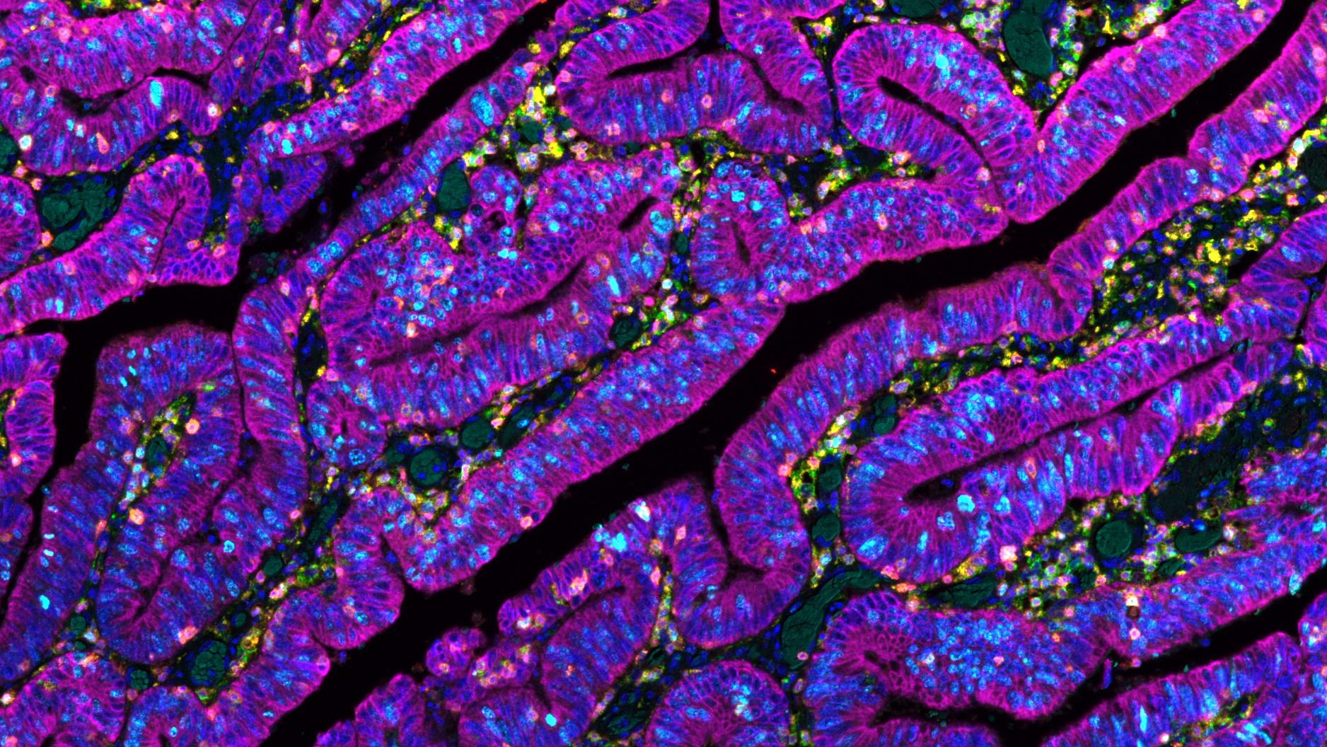
The validation project is scheduled to begin in November.
From Concept Life Sciences
“The Spatial Biology team is thrilled to begin working closely with the Centre for Human Specific Research. Their scientific guidance and funding have been instrumental in enabling us to explore antibody mimetics for those targets which are difficult to detect, or for which reliable antibodies are not available. I look forward to seeing the results of this exciting partnership.”
Dr Giulia Tagliavini; Senior Scientist in the Spatial Biology Team
“This collaboration reflects our commitment to advancing cutting-edge, sustainable technologies that elevate research precision and efficiency – driving progress through smarter, future-ready solutions.”
Dr Hayley Gooding; Biology Services Director