
The Centre for Human Specific Research is collaborating with the Spatial Biology team at Concept Life Sciences to validate two antibody mimetics as replacements for animal-derived antibodies in automated immunohistochemistry (IHC) staining. In this second and final project update, we outline the outcomes of the concluding phases of the Transition Consultancy, which focus on the validation work.
Recap
Concept Life Sciences identified performance issues with a small number of key antibody-based IHC staining assays when applied to disease tissue samples, including poor staining quality and high background signal. The Transition Consultancy focused on validating antibody mimetics as an alternative. Mimetics have the potential to overcome common antibody limitations, such as high background staining, while also avoiding the use of animals or animal-derived products (ADPs) in their development. This reduces ethical concerns and minimises manufacturing issues such as batch-to-batch variability. Furthermore, due to their molecular design, no blocking step is required before application, removing the need for traditional blocking solutions that often contain ADPs. This streamlines assays, shortens processing time, and could lower overall costs by eliminating blocking reagents altogether.
Funding Support for Validation
To begin the validation process, we funded the purchase of a PD-L1 Ankyron (V5, His-tagged; clone: 01S20140), which had been internally validated by ProImmune for use in immunohistochemistry (IHC), alongside an anti-V5 secondary reagent (HRP). All additional reagents and materials were provided by Concept Life Sciences. Staining was performed on formalin-fixed paraffin-embedded (FFPE) cancer tissue known to express PD-L1 and comparisons were made between the performance of the Ankyron and a previously used PD-L1 antibody.
Staining was carried out using the BOND Polymer Refine Detection Kit DAB on a BOND RX autostainer, following protocols recommended by ProImmune. Concept Life Sciences used the suggested Ankyron and secondary reagent concentrations, together with the recommended antigen retrieval conditions, to ensure consistency throughout the validation process (Table 1).
Table 1. Overview of staining conditions.
| Marker | Antigen Retrieval Buffer | Dilution | Secondary Reagent | Dilution |
|---|---|---|---|---|
| PD-L1 (Ankyron) (clone: 01S20140) | ER2 (pH9) 10 min | 10 ug/mL | Anti-V5 HRP Tag | 1:500 |
| PD-L1 (Antibody) | ER2 (pH9) 20 min | 1:100 | Lecia Polymer | N/A |
Initial testing on human non-small cell lung cancer (NSCLC) and triple negative breast cancer (TNBC) tissue sections produced some staining with the PD-L1 Ankyron. However, the signal was considerably weaker and less specific than that achieved using the PD-L1 antibody and was notably fainter than the representative images available on the ProImmune website which was performed on FFPE TNBC tissue (Figure 1).
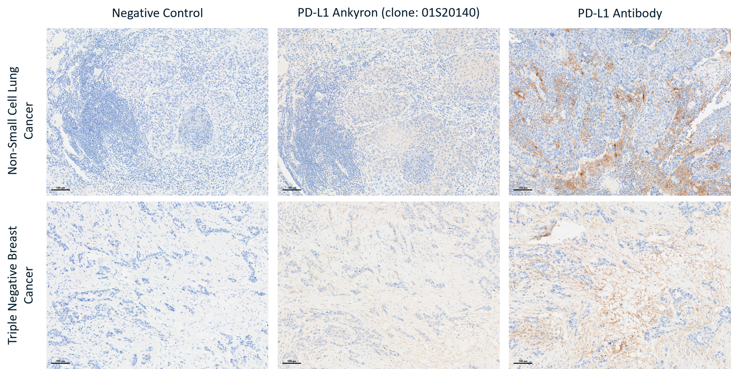
Figure 1. Representative images of DAB staining for PD-L1 Ankyron (clone 01S20140) and PD-L1 antibody in human non-small cell lung cancer and triple negative breast cancer. Scale bar = 100µm.
Following discussion of these results, it was decided that ProImmune would undertake additional in-house validation work on clone 01S20140. In the meantime, they supplied a second internally validated clone, BS40925, free of charge for further testing by Concept Life Sciences, who performed staining using the same staining protocol and conditions as the first validation (Table 2).
Table 2. Overview of staining conditions.
| Marker | Antigen Retrieval Buffer | Dilution | Secondary Reagent | Dilution |
|---|---|---|---|---|
| PD-L1 (Ankyron) (clone: BS40925) | ER2 (pH9) 10 min | 10 ug/mL | Anti-V5 HRP Tag | 1:500 |
| PD-L1 (Antibody) | ER2 (pH9) 20 min | 1:100 | Lecia Polymer | N/A |
When the new clone was evaluated on human NSCLC no detectable staining was observed, with results comparable to the negative control. The PD-L1 antibody showed positive staining pattern consistent with the known expression patterns (Figure 2).
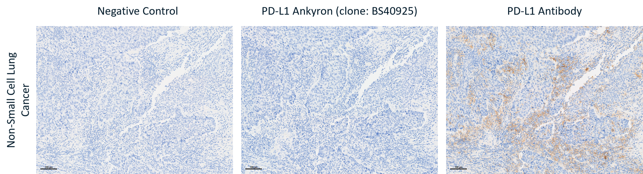
Figure 2. Representative images of DAB staining for PD-L1 Ankyron (clone BS40925) and PD-L1 antibody in human non-small cell lung cancer and triple negative breast cancer. Scale bar = 100µm.
Further discussions lead to the recommendation that extending the antigen retrieval step to 40 minutes could improve staining performance (Table 3).
Table 3. Overview of staining conditions.
| Marker | Antigen Retrieval Buffer | Dilution | Secondary Reagent | Dilution |
|---|---|---|---|---|
| PD-L1 (Ankyron) (clone: 01S20140) | ER2 (pH9) 40 min | 10 ug/mL | Anti-V5 HRP Tag | 1:500 |
| PD-L1 (Ankyron) (clone: BS40925) | ER2 (pH9) 40 min | 10 ug/mL | Anti-V5 HRP Tag | 1:500 |
| PD-L1 (Antibody) | ER2 (pH9) 20 min | 1:100 | Lecia Polymer | N/A |
Under these revised conditions, clone 01S20140 produced faint staining, but the signal still fell far short of the strong staining achieved with the PD-L1 antibody control and was non-specific. Clone BS40925 again showed no staining (Figure 3).
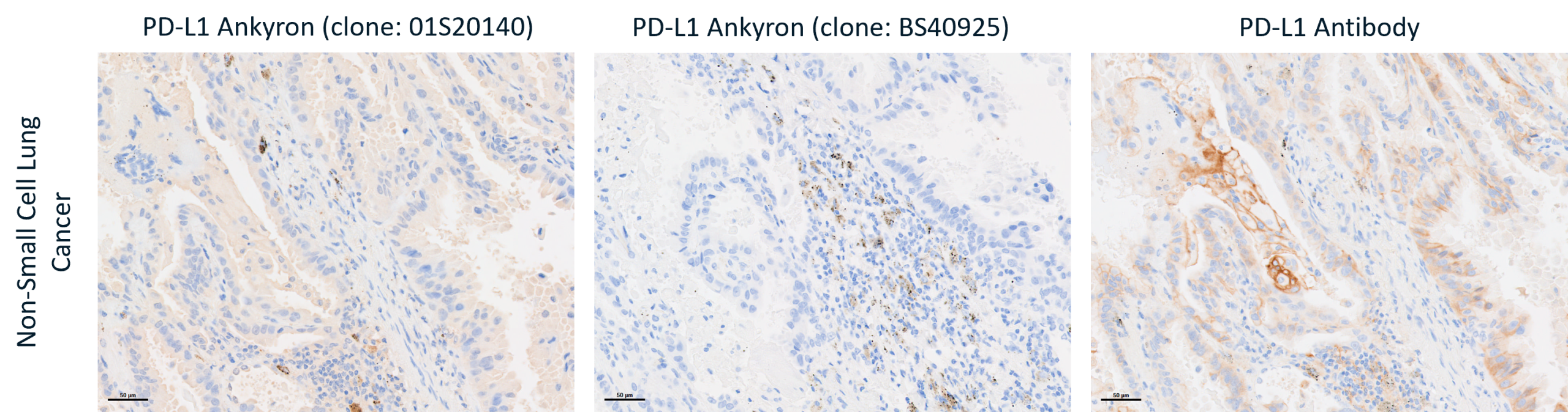
Figure 3. Representative images of DAB staining for PD-L1 Ankyron (clone 01S20140), PD-L1 Ankyron (clone BS40925), and PD-L1 antibody in human non-small cell lung cancer. Scale bar = 50µm.
Based on the results from these initial validation studies, Concept Life Sciences concluded that the PD-L1 Ankyrons could not currently be offered as an alternative to antibody-based FFPE tissue staining using these particular methods and additional validation would fall out with the scope of this project. The decision was taken to not progress further. Although this study did not identify a suitable replacement for the PD-L1 antibody in this application, the project provided valuable insight into the challenges of validating new reagents for automated IHC workflows.
Removing Animal-Derived Products from Final Formulation
Throughout the project, ProImmune demonstrated a willingness to adapt existing products in support of more sustainable, ethically responsible animal-free research practices. Although the Ankyrons provided by ProImmune are entirely animal-free, certain secondary reagents depend on ADPs during manufacturing and final formulation. Over the course of the project, ProImmune implemented measures to address these dependencies, including the introduction of an anti-V5 reagent formulated with an animal-free stabiliser as an alternative to bovine serum albumin (BSA).
Final Thoughts
While the PD-L1 Ankyrons evaluated during the study did not achieve staining performance comparable to established antibody-based methods and were therefore not considered suitable replacements for this application, the project highlighted the importance of rigorous step by step validation when introducing novel reagents into established laboratory processes. Even when protocols and reagents are standardised, factors such as tissue-specific characteristics, inter-laboratory variability, and platform-dependent staining methodologies can influence performance.
Beyond the scope of the initial project, the collaboration encouraged further progress towards reducing reliance on animal-derived products, with ProImmune taking meaningful steps to reformulate supporting reagents using animal-free alternatives.