


As biomedical research increasingly embraces human-specific approaches, new technologies are emerging that have the potential to transform how scientists’ study human biology and disease. At the forefront of this shift are the researchers developing tools that can generate deeper biological insights while improving the efficiency and reproducibility of experiments.
In this article, Giuseppe Di Caprio, Senior Lecturer and Chancellor’s Fellow within the Centre for the Cellular Microenvironment at the University of Strathclyde, shares insights into his journey from optical physics to biomedical engineering and explains how his team is developing a new generation of “smart microscopes” capable of making decisions based on what they see.
At the Interface of Computational and Experimental Biology
“I find it increasingly difficult to separate experimental and computational biology. The two disciplines are most powerful when they are developed together.”
Dr Giuseppe Di Caprio
Microscopy has long been a cornerstone of biomedical research, enabling scientists to visualise cells and tissues in remarkable detail. However, for many researchers it remains a time-consuming and highly specialised process. Critical decisions, such as how samples are prepared, which regions of interest are selected, and when additional data are collected, often rely heavily on the experience and judgement of individual scientists.
This heavy reliance on manual decision-making has led Dr Giuseppe Di Caprio to ask a simple but ambitious question: can microscopy be made smarter?
“My interdisciplinary background has strongly shaped how I approach biomedical research: I am interested not simply in developing new technologies, but in understanding how those technologies can provide better ways of addressing important biological and medical questions.”
Dr Giuseppe Di Caprio
With a background in optical physics, Dr Di Caprios’s research career has centred largely on developing technologies that combine microscopy, microfluidics, and computational approaches for the characterisation of biological systems.
Many of his current research interests can be traced back to his time in Boston. After completing a PhD, he spent more than a decade working alongside researchers in a medical school with a strong focus on imaging and image analysis. There, he saw first-hand the wealth of biological information contained within microscopy data. Yet much of this information remained untapped, with researchers only beginning to explore how imaging data could be analysed quantitatively to reveal deeper insights into cellular behaviour.
“During my time in Boston, I became increasingly interested in the idea that microscopy could be used not just to produce images, but as a quantitative measurement technology.“
Dr Giuseppe Di Caprio
This idea has since become a central theme throughout his research. Dr Di Caprio believes that cellular morphology and dynamics contain valuable information about cellular function, and that rigorous quantitative analysis, supported by machine learning, can provide insights that complement and, in some situations, rival more conventional biochemical assays.
The challenge is creating tools that can reliably extract this information at scale.
Building a Smart Microscope
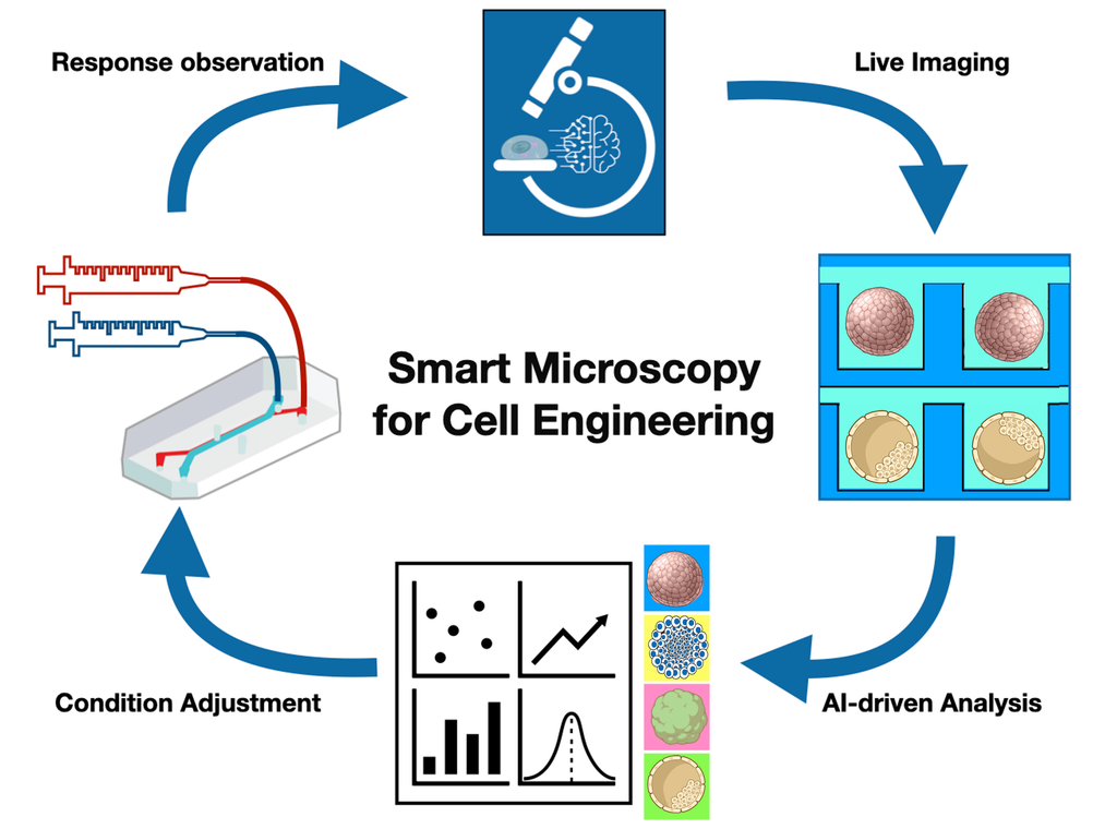
At the heart of Dr Di Caprio’s current research is the development of a “smart microscope”, an imaging system that can analyse what it sees and adapt its behaviour accordingly.
“The key difference from a conventional microscope is that this process happens continuously, creating a feedback loop between observation and action. The image is therefore not simply the final output of the experiment, but it becomes an input that helps determine what the microscope does next.“
Dr Giuseppe Di Caprio
By reducing reliance on individual expertise, Dr Di Caprio believes smart microscopy could help standardise experimental workflows and improve reproducibility. At the same time, it could enable researchers to collect richer datasets that better capture biological variability and strengthen the statistical power of experiments.
“I hope smart microscopy will ultimately relieve scientists from some of the most formulaic and repetitive components of their work, allowing them to spend more time on the aspects of research that require scientific judgement, creativity, and interpretation.“
Dr Giuseppe Di Caprio
How Does It Work?
A smart microscope combines three core components: imaging hardware to capture data, computational algorithms to interpret what is being observed, and automated control systems that can respond to those observations by adjusting the experiment.
One example is the detection of mitosis, the process of cell division. As these events are relatively rare, continuously imaging cells at high speed and high illumination would be inefficient and could potentially damage the sample.
Instead, a smart microscope can monitor a large area using low-intensity imaging while machine-learning algorithms analyse the images in real time, searching for subtle changes in cell shape that may indicate the onset of cell division.
When a potential event is detected, the microscope automatically adjusts its settings, moves to the relevant location and captures detailed images of the process. Once complete, it returns to low-intensity monitoring, ready to identify the next event.
“The system is not simply automating image acquisition, but it is using what it observes to decide what to measure next.“
Dr Giuseppe Di Caprio
A Move Toward Autonomous and Data-Driven Experimentation
Dr Di Caprio and his team are currently applying these concepts to areas including automated cell-engineering protocols, bone tissue generation for grafting applications, and the evaluation of paracrine therapies aimed at repairing cardiac tissue following hypoxic injury.
Currently, the system can continuously monitor biological processes and use quantitative measurements of cell behaviour and morphology to inform how an experiment progresses.
“If we can make these processes sufficiently robust, imaging could become an alternative or complement to more conventional biochemical assays for monitoring biological state. Instead of taking a sample at discrete time points and performing a destructive endpoint measurement, we can potentially observe the same system continuously and non-invasively, extracting quantitative information about how it evolves over time.“
Dr Giuseppe Di Caprio
The next challenge is closing the loop completely. This means developing computational models capable of reliably identifying important biological states in real time and linking those observations to experimental interventions. To achieve this the team are currently working on the development of the actuation and sample-handling systems.
Ultimately, the goal is a system that can detect a biological event, decide on an appropriate action, and automatically perform the next experimental step without human intervention.
“I would not describe any one of these as an insurmountable scientific challenge. One of the advantages of working on a highly interdisciplinary problem is that there are usually several possible routes to a solution.“
Dr Giuseppe Di Caprio
If realised, such systems could be applied across a wide range of human-focused models, from conventional cell culture through to organoids and organ-on-chip platforms.
“If we can combine quantitative imaging, controlled experimental environments such as microfluidic systems, and computational models, we can begin to build increasingly predictive models of human biology based on human cells and tissues rather than relying on animal models.“
Dr Giuseppe Di Caprio
Expert Advice
For researchers interested in developing their own advanced microscopy systems, Dr Di Caprio emphasises the value of engaging with the growing open-source microscopy community.
“One of the things I find particularly encouraging about this field is how much expertise is shared openly, making it possible for researchers to learn not only about microscopy, but also about building microscope systems, developing actuation protocols, and implementing computational approaches for smart microscopy.“
Dr Giuseppe Di Caprio
He points to communities such as the MicroList Forum, Image.sc and OpenFlexure as valuable resources for anyone looking to learn more about microscopy, image analysis, and microscope engineering.
“Many of the challenges involved in building these systems have already been encountered and solved by someone else. I am also always happy to have a conversation with researchers who are interested in these approaches, and to help where my experience might be useful.“
Dr Giuseppe Di Caprio
Final Thoughts
As biomedical research becomes increasingly data-driven, technologies that can continuously observe, interpret and respond to biological systems have the potential to transform how experiments are conducted. For Dr Di Caprio, the smart microscope represents more than an advance in imaging technology. It reflects a broader shift towards integrating computational and experimental biology to create more informative, reproducible and predictive research models.
While some challenges remain, progress in imaging, machine learning, and automation is steadily bringing this vision closer to reality. As these capabilities mature, smart microscopy could help researchers extract more information from human cells and tissues while reducing reliance on labour-intensive and highly specialised workflows.
Ultimately, by combining quantitative imaging with advanced computational models, smart microscopes could become an important part of the toolkit supporting the development of more human-relevant approaches to studying biology and disease.